Understanding Pneumatic Otoscopy
Pneumatic otoscopes add insufflation capability to standard otoscopes, allowing clinicians to assess tympanic membrane mobility—a critical diagnostic technique for identifying middle ear effusion, perforation, and other conditions. The Welch Allyn pneumatic otoscope system integrates a sealed specula system with an insufflator bulb attachment.
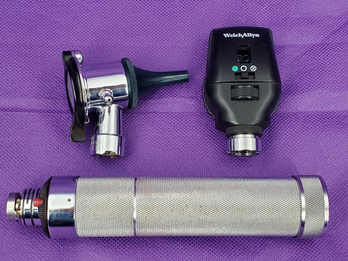
System Components
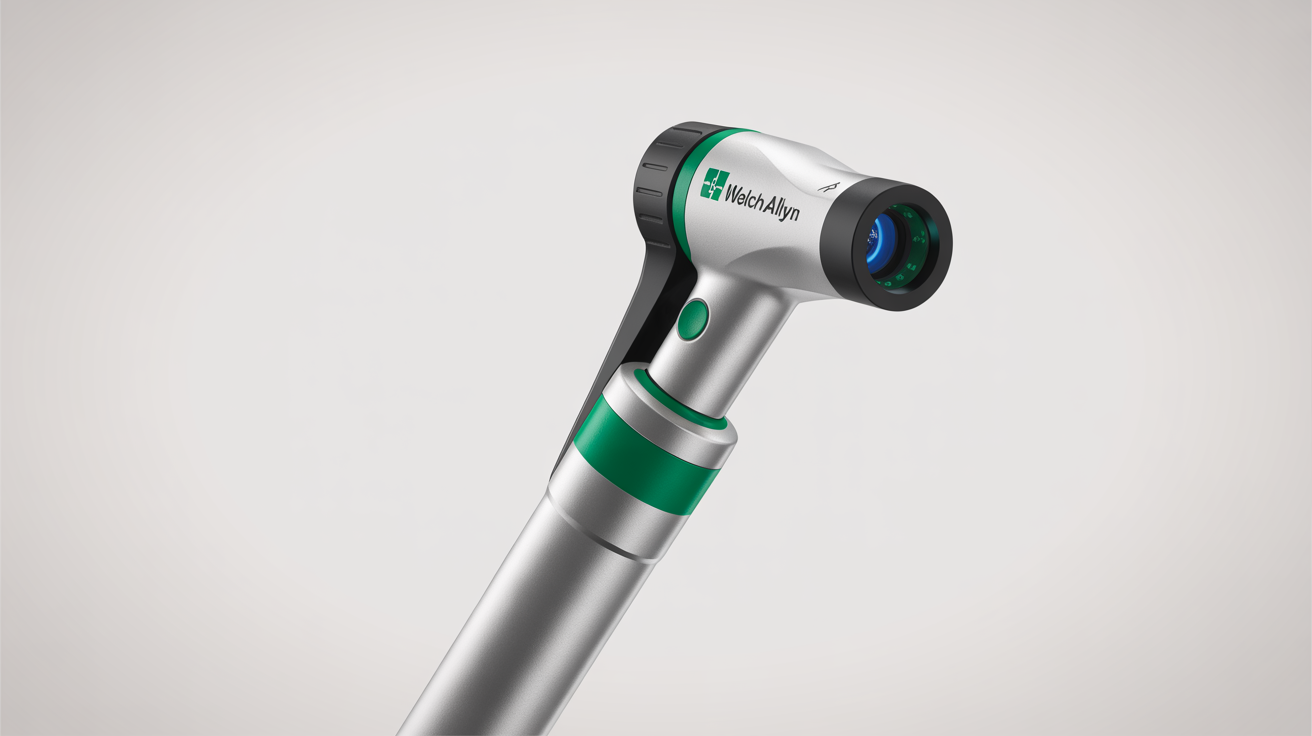
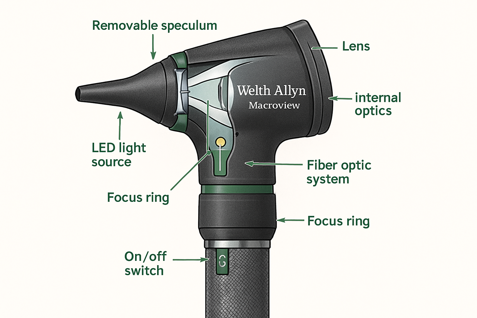
- Otoscope Head: Standard MacroView or conventional optical head with sealed specula port
- Pneumatic Specula: Specialized specula with rubber seal for airtight fit in ear canal
- Insufflator Bulb: Hand-operated bulb generating positive and negative pressure
- Tubing Assembly: Connects insufflator to otoscope head
Diagnostic Technique
Proper Technique for Pneumatic Otoscopy
- Select appropriately sized pneumatic speculum for patient's ear canal
- Attach insufflator bulb to otoscope via tubing connection
- Insert speculum to create gentle seal in ear canal
- Visualize tympanic membrane under illumination
- Gently squeeze insufflator bulb to create positive pressure
- Observe tympanic membrane movement (normal: visible inward deflection)
- Release bulb to create relative negative pressure
- Observe return movement (normal: visible outward deflection)
Clinical Interpretation
- Normal: Brisk movement with pressure changes
- Effusion: Reduced or absent movement due to fluid behind membrane
- Perforation: No movement, possible air escape sound
- Scarring/Adhesions: Limited or asymmetric movement
Common Problems and Solutions
Problem: Insufficient Pressure Generation
Causes: Leak in pneumatic system, worn insufflator bulb, poor speculum seal, damaged tubing
Diagnostic Steps:
- Perform leak test: Occlude specula tip, squeeze bulb—should maintain resistance without constant squeezing
- If leaking, systematically test: Bulb integrity (submerge in water while compressed), Tubing connections and condition, Specula seal quality, Otoscope head seal integrity
Resolution: Replace defective components. Insufflator bulbs (Part #: 05054-200) should be replaced annually or when function degrades.
Problem: Specula Will Not Seal in Ear Canal
Causes: Incorrect speculum size, hardened rubber seal, damaged seal, excessive cerumen
Resolution:
- Ensure correct specula size selection (too small won't seal, too large uncomfortable)
- Replace pneumatic specula if seals hardened (typically every 6-12 months)
- Clean ear canal of excessive cerumen before examination
- Consider reusable metal specula with separate rubber seal attachments for better longevity
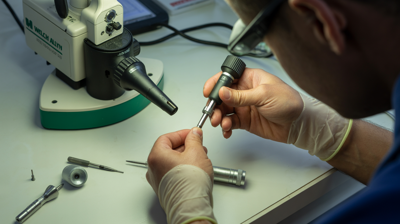
Maintenance Procedures
Daily Maintenance (Clinical Staff)
- Wipe specula and exterior surfaces with disinfectant after each patient
- Inspect pneumatic specula seal for damage
- Verify insufflator bulb function
- Store with tubing unkinked
Weekly Maintenance
- Replace used disposable pneumatic specula stock
- Clean reusable specula per facility sterilization protocol
- Perform leak test to verify system integrity
- Inspect tubing for cracks or degradation
Quarterly Maintenance (Biomedical Engineering)
- Complete functional test of pneumatic system
- Replace insufflator bulb and tubing if showing wear
- Deep clean otoscope head
- Verify optical quality
- Document maintenance activities
Component Specifications
| Component | Part Number | Replacement Interval |
|---|---|---|
| Insufflator Bulb Assembly | 05054-200 | 12 months or when function degrades |
| Pneumatic Specula (2.5mm) | 52334-U | Disposable after use or when seal damaged |
| Pneumatic Specula (4mm) | 52344-U | Disposable after use or when seal damaged |
| Replacement Tubing | 05260-100 | 24 months or when cracking observed |
Infection Control
Critical Considerations
- Pneumatic otoscopy creates direct air pathway from insufflator through ear canal—potential for cross-contamination
- Use disposable pneumatic specula when possible
- If reusable specula used, must undergo high-level disinfection or sterilization
- Insufflator bulb difficult to disinfect internally—consider single-patient use or barrier protection
- Never share pneumatic otoscopes between isolation patients and general population without complete disinfection
Recommended Practices
- Use disposable pneumatic specula for all patients
- Wipe external surfaces of insufflator bulb between patients
- Replace insufflator bulb if internal contamination suspected
- Clean and disinfect tubing per facility protocols
- Consider dedicated devices for isolation areas
Troubleshooting Pneumatic System
Systematic Leak Detection
If pneumatic function inadequate, isolate leak point:
- Test Bulb: Disconnect from system, squeeze underwater—if bubbles appear, replace bulb
- Test Tubing: Block both ends, submerge—if bubbles appear, replace tubing
- Test Speculum Seal: Occlude specula tip with finger, squeeze bulb—if doesn't hold pressure, replace speculum
- Test Otoscope Head: If all above pass but system still leaks, otoscope head pneumatic port seal damaged—requires service
Frequently Asked Questions
⚠ Clinical Safety Considerations
- Never apply excessive pressure—may cause patient pain or tympanic membrane perforation
- Do not perform pneumatic otoscopy if tympanic membrane perforation suspected—air may enter middle ear causing discomfort or spreading infection
- Ensure proper infection control to prevent cross-contamination
- Replace consumables on schedule to maintain function and patient safety
Legal Disclaimer: This guide is for biomedical technicians and trained clinical staff. Pneumatic otoscopy should only be performed by clinicians trained in proper technique. Improper use may cause patient injury.